Norman Chideckel MD Vein Center
212-993-6133

Welcome to the Vascular Surgery and Vein Center

Welcome to the Vascular Surgery and Vein Center, a comprehensive vascular practice providing state-of-the-art care to patients throughout the tri-state area. Under the direction of board-certified surgeon Dr. Norman Chideckel, we utilize our advanced skills and many years of experience to help patients achieve clear, beautiful and healthy legs.

With our convenient Manhattan location, Dr. Chideckel offers patients a wide range of services to treat varicose and spider veins, deep vein thrombosis, and other vascular conditions, including:

Dedicated to helping patients achieve their desired results through the most effective and minimally invasive treatments available, Dr. Chideckel and his staff take the time to talk with each individual patient and address any concerns or questions they may have before undergoing treatment. Your comfort and satisfaction are among our top priorities.

To learn more about the services provided by Dr. Chideckel, please contact us to schedule an appointment. We always welcome new patients and look forward to meeting you.

Vascular Surgery & Vein Center
108 East 96th Street
Front 1
New York, NY 10128
Call us today at 212-993-6133 to schedule an appointment.

We now offer Telemedicine sessions.  Please call us for more information or to schedule a Telemedicine appointment.


See Our Main Website At www.VeinsDoc.com
Blog Posts Are Below:


Category Archives: Venous Ultrasound

Venous Ultrasound

What is venous ultrasound? Vein specialists use venous ultrasound to create images of the body veins. The ultrasound machine emits sound waves and creates vein images. Doctors use this diagnosis procedure to identify blood clots, especially in the leg veins. For instance, when you suffer from deep vein thrombosis, the doctor takes ultrasound images to identify the severity of the condition. Ultrasound is a safe diagnostic procedure because it doesn’t use ionizing radiation. These radiations are harmful to the body.

Venous Ultrasound
(Source)

Before the procedure, the doctor may ask you to avoid drinking and eating for six to eight hours. Also, you should wear comfortable dresses and leave jewelry items at home. The doctor may also ask you to wear a gown.

Venous Ultrasound Imaging – What is it

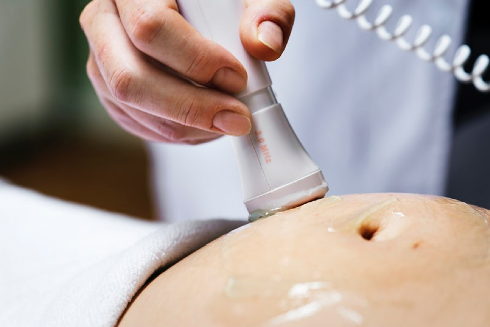
Ultrasound imaging helps vein specialists to diagnose venous and determine effective venous procedure. This is essentially a noninvasive medical procedure that helps doctors to produce pictures of veins through sound waves. The doctor applies gel on the skin and uses a transducer to take the image. The transducer is a small one that releases high-frequency sound waves. The probe collects the sound waves and produces an image on the computer.

The ultrasound machine creates images in real-time. This helps doctors to understand the vein structure. Also, doctors can view blood flow and spot the right position of the disorder. Venous ultrasound creates a picture of veins throughout the body.

The Common Uses of Venous Ultrasound

Vein specialists use ultrasound machines to identify blood clots, especially in the legs. Healthcare providers refer to this procedure as deep vein thrombosis. These blood clots travel throughout the body and block the blood supply to the lungs. These clots can cause life-threatening conditions such as pulmonary embolism. Upon early identification of blood clots, doctors can prevent the clot from passing to the lungs. The healthcare provider performs a venous ultrasound for the following reasons:

  • It identifies long-standing leg swelling and varicose veins. When the valves inside the veins are damaged, the blood pools in the vein. The venous ultrasound identifies the blood pool due to varicose veins. Doctors can identify the problem and remove the veins from the body.
  • Venous ultrasound helps in performing treatment efficiently. While performing invasive procedures, the doctor can check the right position to enter needles and catheters. This helps doctors to prevent bleeding and nerve damage.
  • Healthcare professionals can mark the veins and arteries before performing a procedure. Also, they can identify whether it is safe to perform the procedure or there are complications. For instance, when doctors remove the superficial varicose veins, they should assure the right location for incision.
  • They can assess a blood vessel graft for dialysis, especially when the graft is blocked or narrowed.

The doctor might also use venous ultrasound in children. During congenital vascular malformation and dialysis fistula, the doctor may use venous ultrasound. It helps them identify the connection between a vein and an artery.

While performing a surgery, the doctor places a tiny incision on the children’s leg. This might cause blood clotting due to small vessel size. Also, a blood clot might appear on the arm due to compression. In both conditions, the doctor uses venous ultrasound to evaluate the arteries. Here are some benefits of using venous ultrasound for doctors:

  • Identification of blood clotting
  • Increase in blood flow due to infection
  • Reduced blood flow to various organs
  • Tumor and congenital vascular malformation
  • Narrowing of vessels

Venous Ultrasound | Vascular Surgeon NYC
(Source)

Things to Remember Before the Procedure

Before the procedure you should:

  • Consume every medication prescribed by the doctor
  • Wear comfortable clothes
  • Arrive 15 minutes before the appointment

During Your Exam

When you arrive at the doctor’s clinic, they will ask you to:

  • Lie on the examination table with your hands on the side. The doctor will apply a gel and place the transducer on the veins and arteries they want to capture.
  • When the physician will perform the diagnosis, you might hear the blood pumping throughout the room. You don’t need to be afraid as this is normal.
  • The examination might take up to 15 to 30 minutes.

After Your Exam

After the examination, you can walk and even leave for your home. There isn’t any post-exam instruction. A vein specialist will review your results and identify your health condition.

Venous Ultrasound: Conclusion

A venous ultrasound is a simple and harmless procedure that doesn’t include any side effects. The device uses sound waves to captures the veins. With the help of this diagnosis, vein specialists can identify varicose veins and other venous conditions. Furthermore, they can also spot blood clots and performs procedures to prevent life-threatening conditions.

Do you live in New York and want to consult a vein specialist? If yes, then you should contact the Vascular Surgery and Vein Center in New York. Our experienced vein specialist, Dr. Norman Chideckel treats a wide range of venous diseases. He uses advanced procedures, both non-invasive and invasive to treat conditions such as varicose veins.

For more information, give us a call at 212-993-6133.

Vascular Treatments: Venous Ultrasound

Venous ultrasound produces images of the veins in your body with the help of sound waves. Usually, a doctor uses it to search for blood clots, especially in the veins of the leg. If there are blood clots, the condition is referred to as deep vein thrombosis. Ultrasound has no recognized damaging effects because it does not use ionizing radiation.

Occasionally, your doctor might ask you not to drink or eat anything. You can only have water six to eight hours before surgery. In some cases, you may not require any particular preparation for this procedure. Do not carry any jewelry with you; wear loose and comfortable clothes. You might have to wear a gown before going for surgery.

Vascular Treatments: Venous Ultrasound
(Source)

Ultrasound is painless and safe. Ultrasound imaging is also known as sonography or ultrasound scanning. Ultrasound examinations do not utilize radiation, whereas x-rays do. Because ultrasound captures images in real-time, it can show the movement and structure of your body’s inner organs. Ultrasound can also demonstrate the flow of blood in vessels.

The Procedure of Venous Ultrasound

Venous Ultrasound is also known as a lower extremity duplex scan. A lower extremity duplex scan is a combination of Doppler and sonography. This scan is a noninvasive and precise test that allows doctors to detect venous thrombosis (clots of blood in the veins of your leg), or damaged valves on the surface of the veins that can cause varicose veins. You can always rely on us to perform the procedure in the most reliable manner. Our doctors are licensed and experienced to perform these procedures with the utmost accuracy every time. Take a look at some of the treatments we offer at our clinic.

As stated above, doctors perform ultrasound by using a transducer and place it over the lower extremity. They apply a clear warm gel on a particular body part and move the transducer across the area to visualize the internal images. Usually, they do it with the patient in the supine position, but they can also perform it while the patient is standing. However, the standing position will allow you to have the most precise evaluation of a defective valve in the vein. The images will appear on the screen in real-time so that you and your doctor can review them together.

Vascular Treatments: Venous Ultrasound
(Source)

What Are the Common Uses of This Procedure?

Surgeons commonly use venous ultrasound procedures to detect blood clots, particularly in the veins of your legs. Hence, whether the doctor chooses this method or some other often depends upon the condition of DVT or vein thrombosis. Unfortunately, the clots in the veins of your legs can pass into the lungs, causing a dangerous disease called pulmonary embolism. If you find blood clots in your leg at an early stage and start your treatment, you can prevent it from transferring to the lung.

Moreover, the following are some more uses of a venous ultrasound study;

  • Determine the cause of long-standing swelling in your leg. Many people have a common condition called “varicose veins.” Normally, the valves in the veins that regulate the flow of blood may be damaged. A venous ultrasound can help you identify the unusual blood flow and defective valves.
  • Aid in directing the placement of a catheter or needle into a vein. Through Sonography, a doctor can locate the accurate area of the vein and avoid damage to nerves and nearby arteries.
  • Identify the veins in your leg or arm so that doctor can remove the pieces of the vein and use it to block blood vessels or bypass a narrowed vessel.
  • Inspect a blood vessel graft used for dialysis to know whether the blood vessel is working accurately or not.

Doppler Ultrasound

Doppler ultrasound is a unique ultrasound technique; doctors can us this technique to evaluate the movements of unwanted substances in your body. Doppler ultrasound also allows your doctor to see and examine blood flow through veins and arteries in your body. A physician can use Doppler ultrasound for a variety of conditions. Some of these conditions are as under.

  • Obstructions in blood flow
  • Thinning of vessels
  • Congenital vascular and tumor malformations
  • Absent or reduced blood flow to different organs of the body
  • Blood flow is more significant than before, which might be a symbol of an infection

Consult NYC Vascular Surgeon Norman Chideckel MD for all Vascular Conditions

Even the most harmless conditions can sometimes indicate a serious problem. Not paying attention on time can result in irrevocable damages. You have to make sure that you consult a specialist before your situation shifts to the worse side.

When you need help, you can always call us at 212-993-6133 to book an appointment. Meet Dr. Chideckel to discuss your options and find the best treatment to your problem. You can also book your appointment online